Underwater Near Infrared Imaging of Chlorophyll Fluorescence and Beyond
Written by Barry Hicks
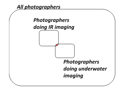
A small fraction of photographers have gone beyond the visible to do digital imaging in the infrared; if you’re on Kolari Visions’ www site you’re probably one of them, or at least, interested in becoming one of them. A small fraction of photographers have gone beyond imaging on land to do digital imaging underwater. A very, very, small fraction of all photographers have begun to do underwater digital imaging in the infrared (red in the Venn Diagram at the right). If you are a scuba diving photographer who is interested in getting started, I invite you to join us by getting your camera converted for IR/full spectrum imaging. If you are an infrared photographer interested in joining us, I encourage you to become scuba certified and join us.
It is “common knowledge” among photographers that there is ample near infrared (NIR) light at the earth’s surface from solar radiation. Like visible light, it can be reflected off of surfaces to allow for NIR digital imaging. It is also “common knowledge” among divers that light penetration is color (wavelength) dependent, and that the longer the wavelength, the less penetration. As red light penetrates less than about 5 m in sea water, it is assumed that NIR light (with an even longer wavelength) penetrates even less. That is accurate as most solar NIR light penetrates less than about 1 meters. But the follow-on assumption that this means NIR light cannot be imaged underwater is not accurate. It is true that you cannot image NIR from as far away as you can on land, but the limitations of NIR underwater imaging have not been fully explored.
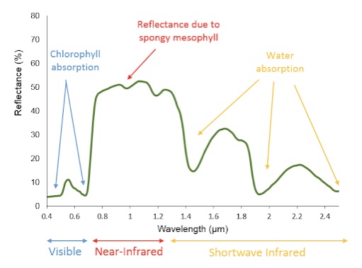
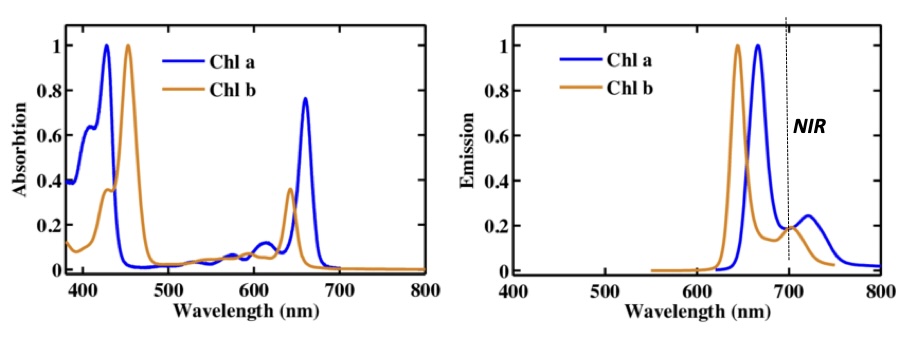
Certainly, if external lighting is supplied, one can image NIR fluorescent emission from any organism that contains chlorophyll. Chlorophyll undergoes a phenomenon known as fluorescence (although sometimes called luminescence in certain fields). This is the application and absorption of relatively high energy light, and emission of relatively low energy light. Unlike bioluminescence or phosphorescence, fluorescent emission only occurs while the specimen is being actively illuminated. Chlorophyll-a and chlorophyll-b absorb both blue and red light (see absorbance spectrum below left). Chlorophyll-a is the common chlorophyll in the terrestrial and the marine environments. By using blue light illumination near 450 nm, both of these can be excited. If you want selective illumination, using 400 nm light for chlorophyll-a and 475 nm light for chlorophyll-b can allow the two to be excited separately. Once chlorophylls (or any fluorescent material) are excited, some of the energy in the exciting photons is lost as heat (kinetic energy). The excited chlorophylls then re-emit light at lower energy (longer wavelength). Thus, the emission (spectrum shown above right) is largely in the red (prominent peak near 650 (b) or 680 (a) nm), but both contain significant emission in the NIR above 700 nm. Since both chlorophylls emit in the NIR, both can be visualized by NIR imaging. This is one reason why chlorophyll-containing subjects (trees, grass, etc.) are often so intensely bright in terrestrial IR images; they are taking in visible light an re-emitting by fluorescence in the NIR portion of the spectrum. Also, chlorophylls tend to reflect NIR light (700-1200 nm or 0.7-1.2 μm) more effectively than visible light (see reflectance spectrum at the right).
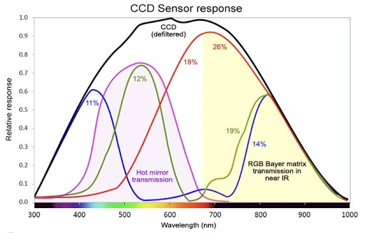
Most common consumer digital cameras these days have complementary metal oxide semiconductor (CMOS) sensors. These sensors actually have good coverage through the NIR, but when you get into the mid IR (above about 1000 nm), they no longer have appreciable sensitivity. A typical spectral response curve for a CMOS sensor is shown (black line), but they vary somewhat by manufacturer. In the commercial cameras, a UV and an IR filter are added (yellow regions) to prevent either of those light sources reaching the sensor. Three different colored bandpass filters are present (a blue pass, a green pass or a red pass) are also present (only one on a given pixel). Above about 725 nm, all three of these filters allow NIR light to pass, albeit not at the same efficiency. By removing the IR filter (yellow right), the camera sensor can detect light even approaching 1000 nm.
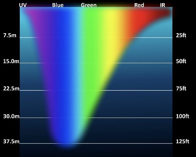
So if NIR light can only travel 1-2 meters in water, how can you do any imaging, especially in water deeper than 5 m? There are two answers to this. First of all, you can do macro photography with external lighting. Macro lens all typically have minimum focal lengths less than 1/10 m and getting as close as possible is usually desirable. From such distances, there is more than enough NIR emitted from chlorophyll containing organisms to image. Be careful, external light sources also put out some NIR light, so those sources must be capped with short pass filters allowing only visible light to reach the target. The second answer is that in the daytime, blue light can penetrate water efficiently even at 30 m (see the distance light travels in sea water at right). Since that light can also excite chlorophyll, it will lead to NIR fluorescent emission from chlorophyll containing specimens. Just how deep chlorophyll NIR fluorescent emission can be detected, as well as how far from an externally illuminated specimen at night NIR emission can be detected are two things we would like to see the underwater NIR diving photographer community investigate. It is interesting to note that some of the marine organisms have chlorophyll pigments in their eyes, even at depths beyond where photosynthesis can occur.
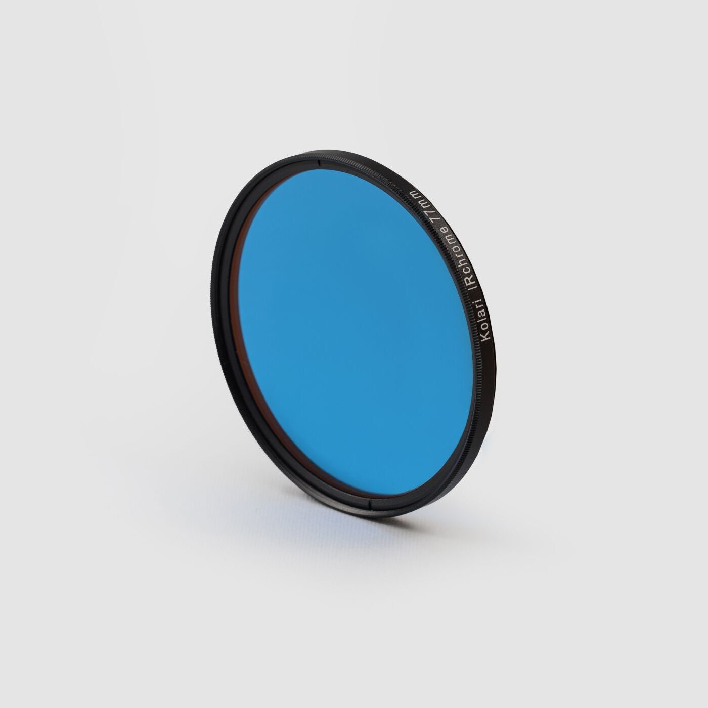
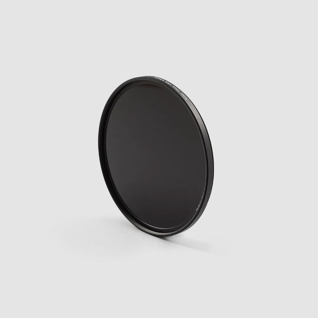
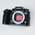
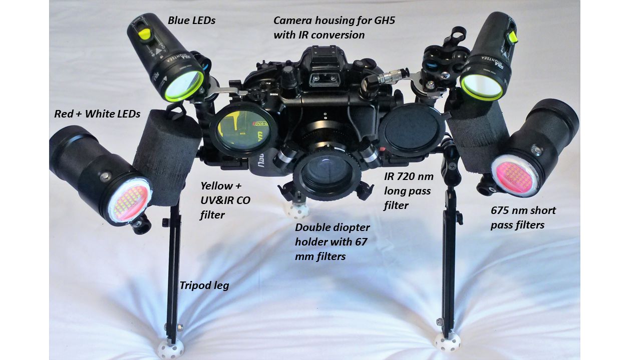
So what all do you need to get started? Basically, an IR sensitive camera, a long pass IR filter, and an underwater housing. First, get a camera upgrade from Kolari (or DIY) so that your camera can image in the NIR. Second, get an underwater housing for your camera. Third, get scuba certified…(actually this isn’t required, but if you want to image corals and related reef critters it would be in your interest). I floated with my camera and housing in the Green River of southwest Wyoming and was able to make videos of NIR chlorophyll fluorescence on the bottom at only about 5 feet depth). If you want to do night work, you’ll need some LED dive lights, and you should put a short pass NIR filter over them to prevent scattered light. Alternatively, if you have IR dive lights, you can image reflectance from them. A picture of our set up is shown above.
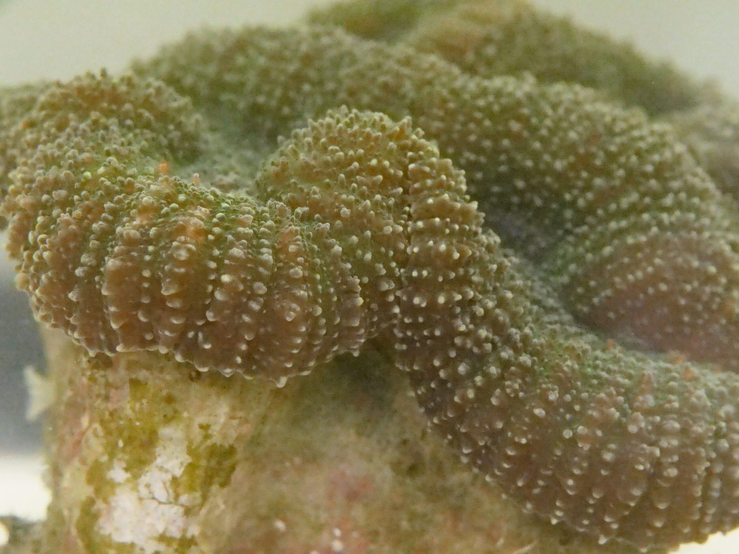
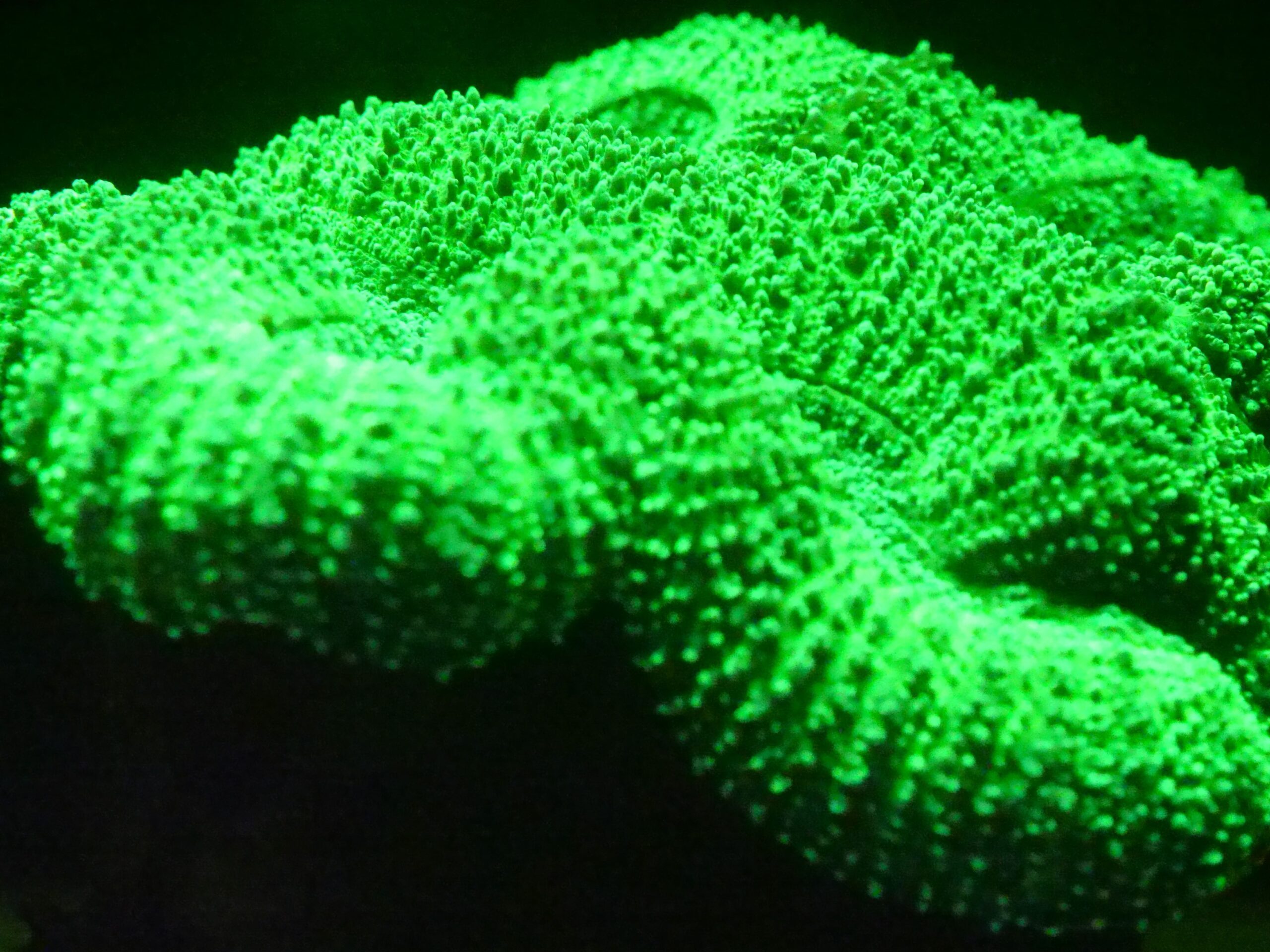
So what would NIR fluorescent images of chlorophyll in underwater wildlife look like? Shown here are three images of an anemone in the Caribbean Sea near Carriacou, Grenada taken with our IR modified Panasonic GH5 camera in a Nauticam housing using two BigBlue 15,000 lumen white LED illumination dive lights with a 675 nm short pass filters on them and a 720 nm NIR long pass filter on the camera. The left most is simply the visible reflectance image (what you see), the center image is the visible fluorescent image from green fluorescent proteins (green) blue LED dive lights and a yellow long pass filter on the camera, and the right most image is of chlorophyll NIR fluorescence, again illuminated with the white LEDs and the 720 nm NIR long pass filter on the camera. This type of imaging was the basis of a scientific paper published in the Journal of Marine Science and Engineering in January 2020. It was also the basis of a Dive Slate article that appeared in Q3 (August) 2020 edition of the magazine Alert Diver.
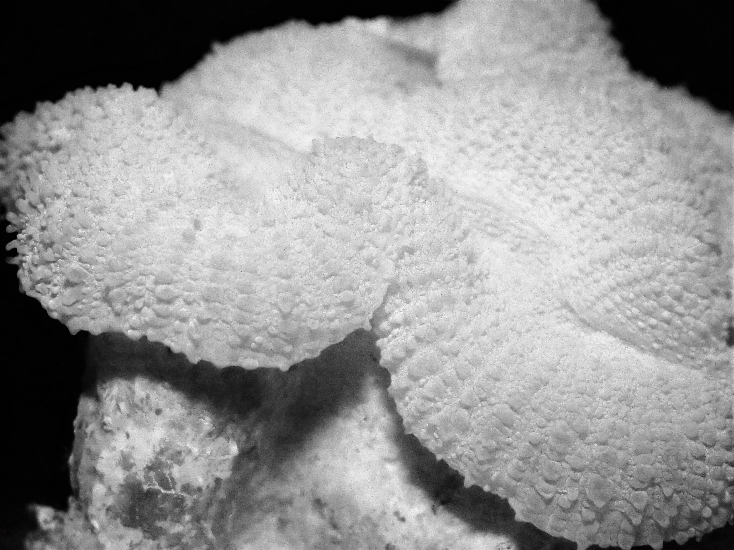
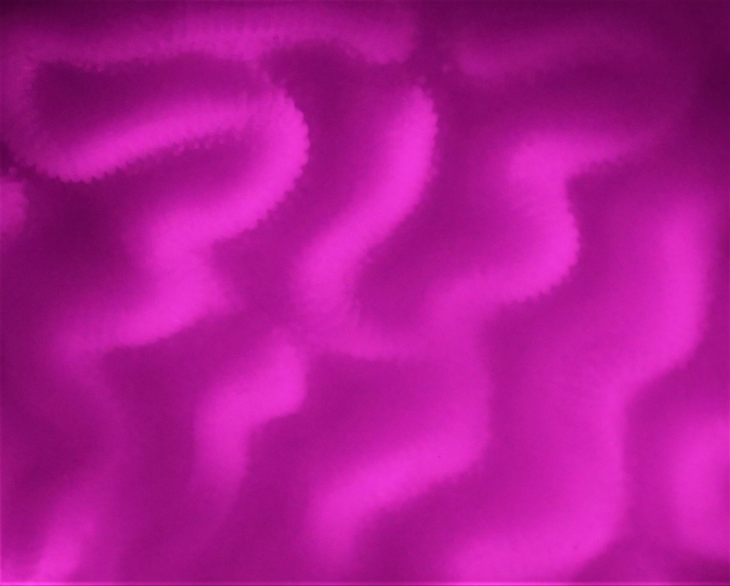
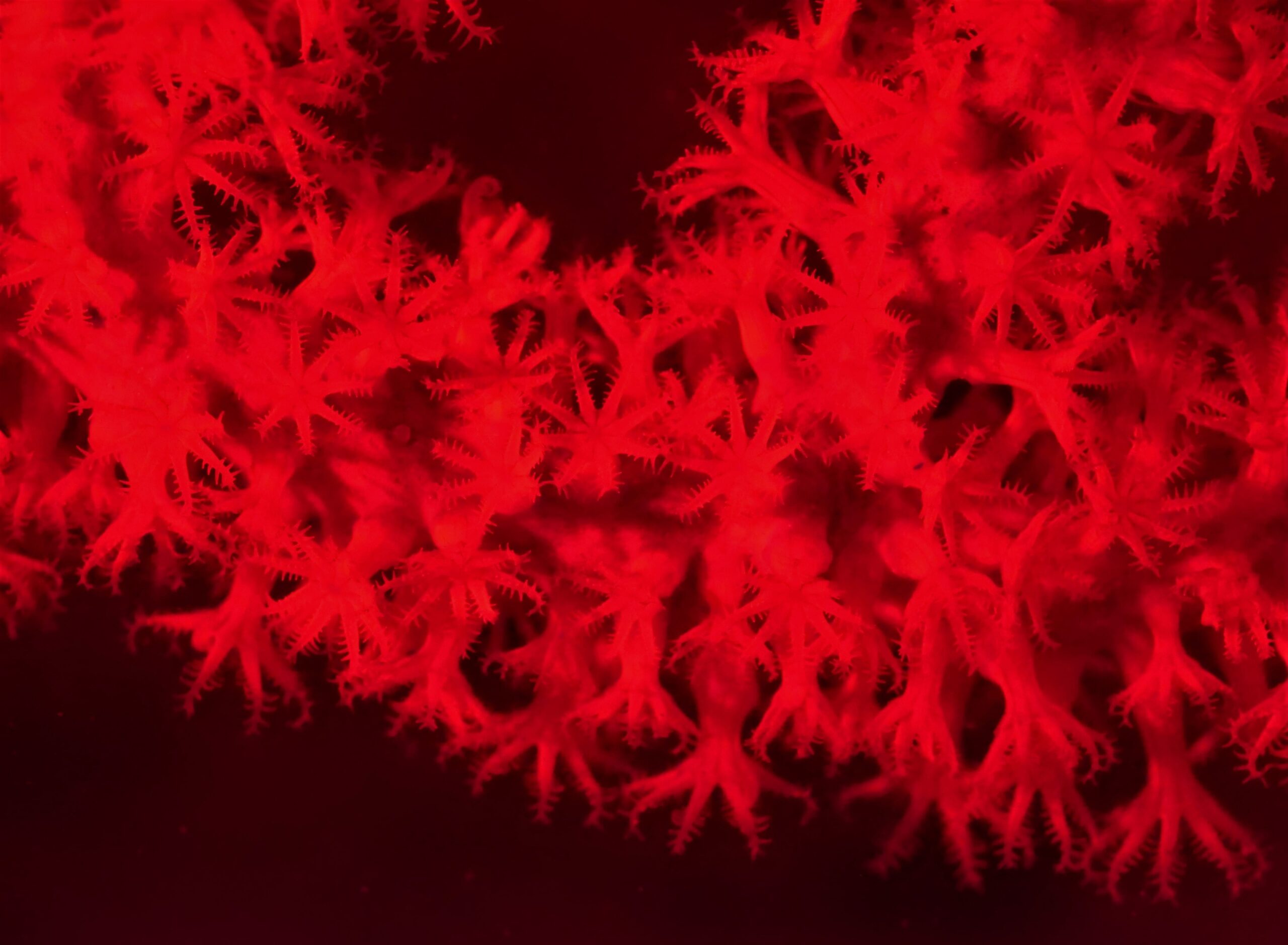
Underwater NIR imaging can also produce artistic effects by altering the white balance setting on the camera, or by using different lighting combinations. Normally, one must use a gray/white slate to set a custom white balance for accurately portraying the difference between background and specimen. This leads to the NIR photos coming out in almost entirely grays as shown for the sea fan above right. But like all photography, the effects one can get by altering lighting, filters and custom white balance settings can be used to significantly alter the results, as shown for the brain coral middle right, or the soft coral below right, so there is always room for artistic involvement. If you want more information, I have a PDF background file giving more description including potential vendors (I have not commercial interests in this!), that is available for free. You can contact me at barry.hicks@afacademy.af.edu.
References
- Mammadov, Gulmammad. Measurement of the Membrane Electric Field and the Swimming Behavior of Chlamydomonas reinhardtii: Experiments and Analytical Modeling. Diss. Syracuse University, 2015.
- Abeer N. Abdul-Hammed. Remote Sensing Optical Images Applications in Vegetation Monitoring and Mapping, Part of Baghdad Supervised by Alaa S. Mahdi. Masters Thesis · September 2022
- Taken from URL: http://www.astrosurf.com/luxorion/Physique/spectral-response-ccd.jpg. Accessed 6 June 2024.
- Taken from URL: https://shops.cheapsalesoutlet2024.ru/content?c=what+color+light+penetrates+water+the+deepest&id=6 . Accessed 6 June 2024.